Subluxing Ecu Tendon refers to the displacement of the Extensor Carpi Ulnaris (ECU) tendon from its normal position, leading to ulnar-sided wrist pain and instability; CAR-DIAGNOSTIC-TOOL.EDU.VN provides comprehensive diagnostic tools and repair guidance to address ECU tendon subluxation effectively. Through advanced diagnostic support and expert repair insights, CAR-DIAGNOSTIC-TOOL.EDU.VN empowers technicians with necessary knowledge and tools, supplemented by training programs and remote expert support to help navigate complex ECU tendon issues and improve diagnostic precision.
Contents
- 1. Understanding Subluxing ECU Tendon: An Expert’s Overview
- 1.1. What are the Key Anatomical Aspects of the ECU Tendon?
- 1.2. What Triggers ECU Tendon Subluxation?
- 1.3. How is Subsheath Tear Classified?
- 2. Diagnosing Subluxing ECU Tendon: Methods and Techniques
- 2.1. What are the Key Physical Examination Techniques?
- 2.2. What Imaging Techniques are Recommended?
- 2.3. What Tools Does CAR-DIAGNOSTIC-TOOL.EDU.VN Recommend for ECU Diagnosis?
- 3. Treatment Options for Subluxing ECU Tendon: A Detailed Guide
- 3.1. When is Conservative Management Recommended?
- 3.2. What Surgical Options are Available?
- 3.3. What is the Surgical Technique for Subsheath Reconstruction?
- 3.4. What Post-operative Care is Required?
- 3.5. What Support Does CAR-DIAGNOSTIC-TOOL.EDU.VN Offer for ECU Treatment?
- 4. Surgical Reconstruction Techniques: A Step-by-Step Approach
- 4.1. What are the Preliminary Steps?
- 4.2. How is the ECU Tendon Exposed?
- 4.3. How is the Extensor Retinaculum Flap Created?
- 4.4. How is the Subsheath Reconstructed?
- 4.5. What are Additional Considerations?
- 4.6. What Resources Does CAR-DIAGNOSTIC-TOOL.EDU.VN Provide for Surgical Guidance?
- 5. Post-operative Rehabilitation: Regaining Wrist Function
- 5.1. What is the Initial Post-operative Phase?
- 5.2. What Happens During the Intermediate Rehabilitation Phase?
- 5.3. What Exercises are Recommended During the Advanced Strengthening Phase?
- 5.4. What are the Guidelines for Returning to Activity?
- 5.5. How Does CAR-DIAGNOSTIC-TOOL.EDU.VN Support Rehabilitation?
- 6. Addressing Post-Surgical Complications: Prevention and Management
- 6.1. What are Common Post-Surgical Complications?
- 6.2. How Can Infections be Prevented and Managed?
- 6.3. How Can Nerve Injuries be Prevented and Managed?
- 6.4. How Can Tendonitis be Prevented and Managed?
- 6.5. How Can Stiffness be Prevented and Managed?
- 6.6. How Can Recurrent Subluxation be Prevented and Managed?
- 6.7. What Support Does CAR-DIAGNOSTIC-TOOL.EDU.VN Offer for Complication Management?
- 7. Emerging Research and Techniques in ECU Tendon Management
- 7.1. What are the Current Research Trends?
- 7.2. What Innovations are There in Imaging Techniques?
- 7.3. What are the Surgical Innovations Being Researched?
- 7.4. What are the Findings of Biomechanical Studies?
- 7.5. How Does CAR-DIAGNOSTIC-TOOL.EDU.VN Incorporate Emerging Techniques?
- 8. Long-Term Outcomes and Patient Satisfaction: What to Expect
- 8.1. What are the Long-Term Results of Subsheath Reconstruction?
- 8.2. What do Studies Say About Patient Satisfaction?
- 8.3. What Factors Influence Long-Term Outcomes?
- 8.4. What Considerations Are Important for Patient Expectations?
- 8.5. How Does CAR-DIAGNOSTIC-TOOL.EDU.VN Enhance Long-Term Outcomes?
- 9. Optimizing ECU Tendon Healing: Nutrition and Lifestyle Factors
- 9.1. What Role Does Nutrition Play in Tendon Healing?
- 9.2. What Foods Support Tendon Healing?
- 9.3. What Lifestyle Factors Impact Healing?
- 9.4. What are the Recommendations for Smoking and Alcohol Consumption?
- 9.5. What are the Guidelines for Hydration and Rest?
- 9.6. How Does CAR-DIAGNOSTIC-TOOL.EDU.VN Support Optimized Healing?
- 10. FAQ: Addressing Common Questions About Subluxing ECU Tendon
- 10.1. What is the Main Cause of ECU Tendon Subluxation?
- 10.2. What are the Symptoms of Subluxing ECU Tendon?
- 10.3. How is ECU Tendon Subluxation Diagnosed?
- 10.4. Can ECU Tendon Subluxation Heal on Its Own?
- 10.5. What is the Conservative Treatment for ECU Tendon Subluxation?
- 10.6. When is Surgery Necessary for ECU Tendon Subluxation?
- 10.7. What are the Surgical Options for ECU Tendon Subluxation?
- 10.8. What is the Recovery Time After ECU Tendon Surgery?
- 10.9. What Activities Should be Avoided After ECU Tendon Surgery?
- 10.10. What Resources Does CAR-DIAGNOSTIC-TOOL.EDU.VN Offer for ECU Tendon Management?
1. Understanding Subluxing ECU Tendon: An Expert’s Overview
Is the subluxing ECU tendon causing you ulnar-sided wrist pain? Subluxing ECU tendon involves the displacement of the Extensor Carpi Ulnaris tendon from its designated groove, resulting in pain and potential wrist instability. This condition, predominantly affecting athletes and individuals with repetitive wrist movements, demands a comprehensive understanding for accurate diagnosis and effective management.
The Extensor Carpi Ulnaris (ECU) tendon is crucial for wrist stability and movement, particularly in activities involving forearm rotation and wrist deviation. Its primary role is to stabilize the wrist during movements, especially those that combine supination, flexion, and ulnar deviation. This function is supported by the ECU subsheath, a fibrous structure that keeps the tendon in place within the ulnar groove.
Damage to the subsheath, often resulting from acute injuries or repetitive stress, can lead to ECU tendon subluxation. This occurs when the tendon slips out of its normal position, causing pain, clicking, or snapping sensations on the ulnar side of the wrist. In chronic cases, this can lead to persistent wrist instability and discomfort.
1.1. What are the Key Anatomical Aspects of the ECU Tendon?
The ECU tendon originates from the lateral epicondyle of the humerus and inserts into the base of the fifth metacarpal. At the wrist, it travels within the sixth dorsal compartment, stabilized by a distinct subsheath. This subsheath acts as a labrum along the ulnar side of the groove, preventing tendon subluxation. The linea jugata, a sling of collagen fibers that inserts onto the interosseus membrane, reinforces it.
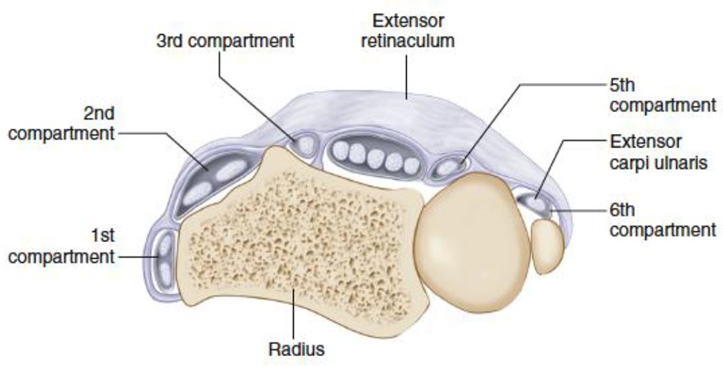 Anatomical illustration of the Extensor Carpi Ulnaris (ECU) tendon within the sixth dorsal compartment of the wrist
1.2. What Triggers ECU Tendon Subluxation?
ECU tendon subluxation occurs when the tendon slips out of its groove. The ECU tendon is under maximum stress when the wrist is supinated, flexed, and ulnarly deviated. Sudden, forceful contraction of the ECU or repetitive minor trauma causes attenuation of both the tendon and the subsheath. Tears to the subsheath are classified into three types: Type A, Type B, and Type C.
1.3. How is Subsheath Tear Classified?
Tears to the subsheath are classified into three types:
- Type A: Occurs at the ulnar side of the subsheath. The tendon can return to the ulnar groove under the torn edge of the subsheath.
- Type B: Occurs at the radial side of the sheath. These are less likely to heal as the tendon lies outside of the torn sheath.
- Type C: Occurs when the fibrous subsheath is detached from the ulna’s periosteum. With this widened subsheath, the tendon can move out of the ulnar groove and remain in a false sheath.
Understanding the classification of subsheath tears is important when evaluating a new patient, as some tears are unlikely to heal with conservative management.
2. Diagnosing Subluxing ECU Tendon: Methods and Techniques
How do you diagnose subluxing ECU tendon effectively? Diagnosing subluxing ECU tendon involves a combination of physical examination techniques and imaging studies to confirm the condition and rule out other potential causes of wrist pain. CAR-DIAGNOSTIC-TOOL.EDU.VN emphasizes the use of accurate diagnostic methods to ensure effective treatment.
2.1. What are the Key Physical Examination Techniques?
A thorough physical examination is crucial in diagnosing ECU subluxation. Key tests include the ECU synergy test and the ECU subluxation test.
- ECU Synergy Test: The patient supinates the wrist, and the examiner applies resistance at the radial side of the hand. Pain during this test indicates constrained ECU tendinopathy.
- ECU Subluxation Test: The patient supinates while the examiner applies resistance to the ulnar aspect of the hand, then ulnarly deviates the wrist. This test can reveal tendon subluxation.
- Heart-Like Test: The backs of the hands are placed together against the chest with the thumbs pointed up. This provoking maneuver can produce a snapping sound and sensation.
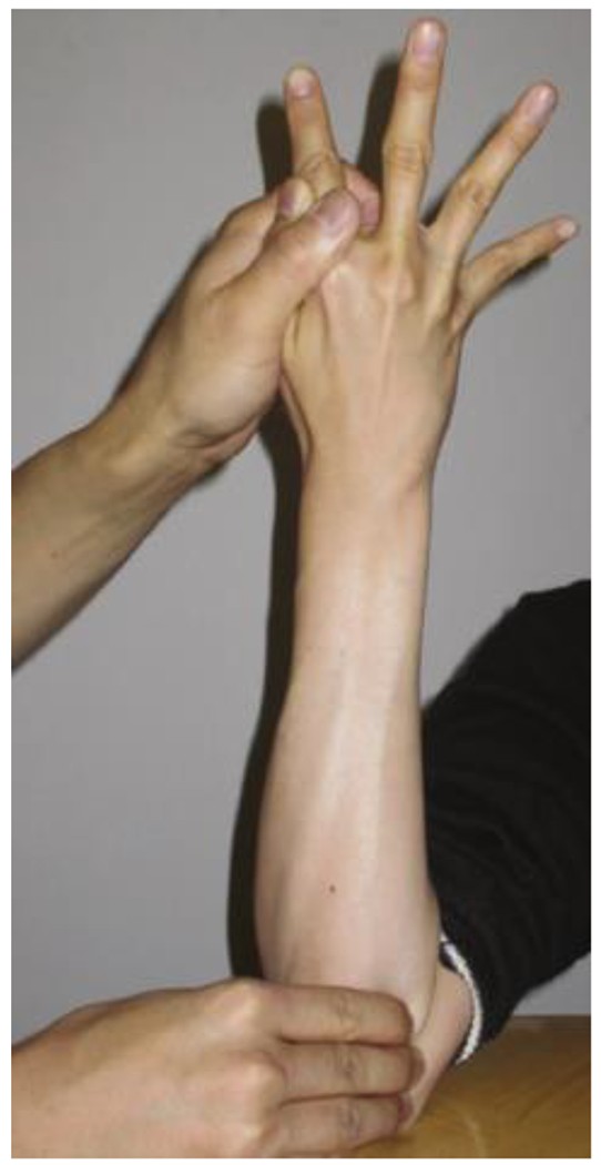 Illustration demonstrating the Extensor Carpi Ulnaris (ECU) synergy test for diagnosing tendon abnormalities
According to a study by Sato et al., the ECU synergy test has a sensitivity of 74% and a specificity of 86% in predicting ECU tendon abnormalities when compared to sonographic evaluation.
2.2. What Imaging Techniques are Recommended?
Imaging techniques play a vital role in confirming the diagnosis and assessing the extent of the injury.
- Wrist Radiographs: Mandatory to rule out bony pathology.
- Ultrasonography: Assesses the ECU tendon due to its superficial location. It identifies ECU subsheath instability, though it can also show instability in asymptomatic patients.
- MRI: Characterizes the anatomy of the tendon and the ulnar groove but is not routine in pre-operative planning.
2.3. What Tools Does CAR-DIAGNOSTIC-TOOL.EDU.VN Recommend for ECU Diagnosis?
| Tool | Use | Benefits |
|---|---|---|
| Diagnostic Ultrasound | Real-time imaging of the ECU tendon and subsheath. | Non-invasive, dynamic assessment of tendon movement, high sensitivity. |
| High-Resolution MRI | Detailed visualization of soft tissues and bony structures. | Provides a comprehensive view of tendon integrity, subsheath condition, and potential associated pathologies. |
| Physical Examination Tools | Goniometer, dynamometer, and palpation aids. | Aids in accurate assessment of wrist range of motion, strength, and tenderness. |
| Diagnostic Software | Tools for analyzing imaging data and generating detailed reports. | Enhances diagnostic accuracy and streamlines the reporting process. |
By integrating these diagnostic approaches, healthcare professionals can accurately identify subluxing ECU tendon and tailor appropriate treatment plans to optimize patient outcomes.
3. Treatment Options for Subluxing ECU Tendon: A Detailed Guide
What are the most effective treatment options for subluxing ECU tendon? Treatment for subluxing ECU tendon ranges from conservative management to surgical interventions, depending on the severity and chronicity of the condition. CAR-DIAGNOSTIC-TOOL.EDU.VN provides insights into both non-surgical and surgical approaches to ensure comprehensive care.
3.1. When is Conservative Management Recommended?
Initial management typically involves conservative measures, especially for acute injuries.
- Immobilization: A long-arm cast for six weeks can help stabilize the wrist and allow the tendon and subsheath to heal.
- Physical Therapy: Exercises to strengthen the wrist and forearm muscles, improve range of motion, and reduce pain.
- Activity Modification: Avoiding activities that exacerbate the condition.
3.2. What Surgical Options are Available?
Surgery is considered when conservative measures fail to provide relief or in cases of chronic instability.
- Primary Subsheath Repair: In acute cases (Type A and B tears), the torn subsheath can be debrided and repaired. For acute Type C tears, the avulsed periosteum is reattached to close the sheath.
- Subsheath Reconstruction: For chronic injuries, reconstruction of the subsheath is often necessary. Techniques involve using flaps of extensor retinaculum with or without bone anchors. The Spinner and Kaplan technique, which uses an ulnarly-based flap to create a sling for the ECU tendon, is a preferred method.
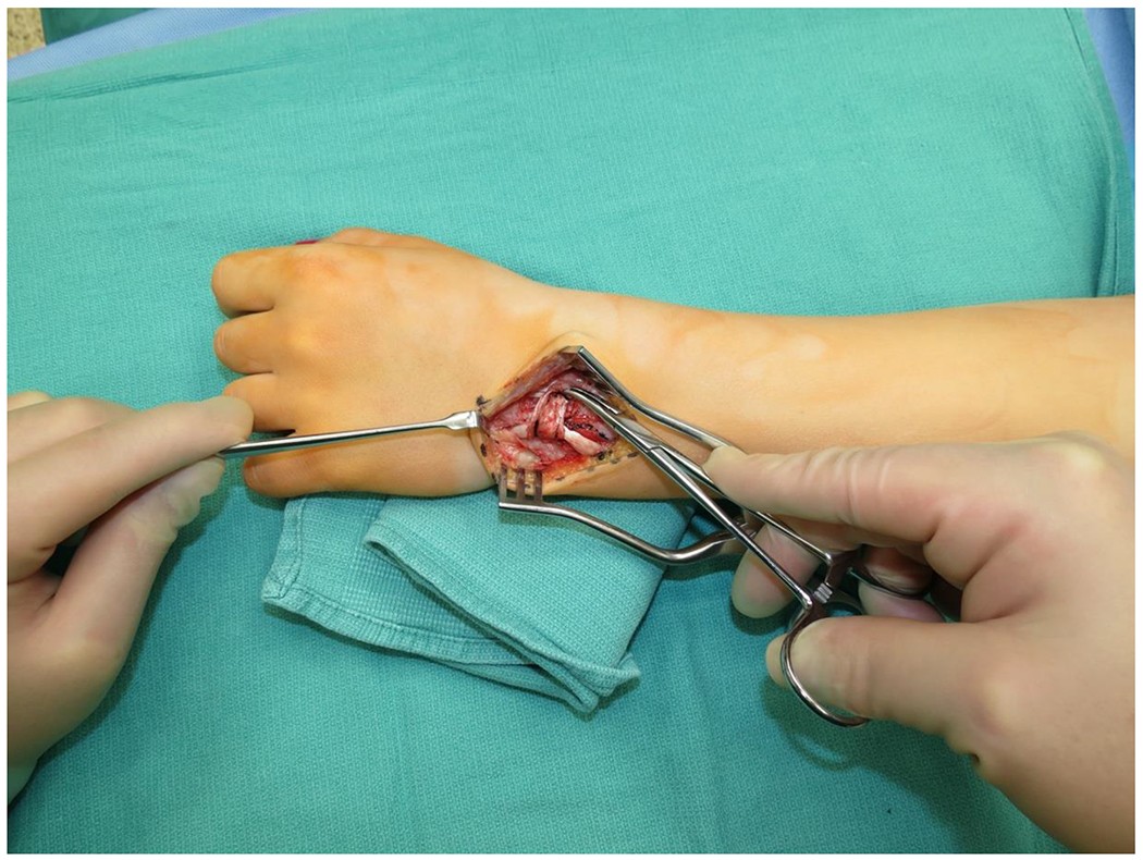 Illustration showing the surgical technique of passing the extensor retinaculum flap under and over the Extensor Carpi Ulnaris (ECU) tendon during reconstruction
3.3. What is the Surgical Technique for Subsheath Reconstruction?
The surgery is performed under general anesthesia or an axillary nerve block with a tourniquet. A five-centimeter incision over the dorsal ulnar wrist is made for exposure. The dorsal sensory branch of the ulnar nerve runs in the subcutaneous tissue in this area and must be avoided. The extensor retinaculum over the 6th compartment is incised to expose the ECU tendon. The torn edges of the subsheath and any frayed tendon are debrided. If the subsheath edges come together without tension, a primary repair can be performed at this stage.
If proceeding with subsheath reconstruction, a 3-cm wide flap is planned. The flap is based at the ulnar border of the fifth extensor compartment. The radial limit is the radial aspect of the third compartment. The flap is carefully elevated from radial to ulnar. The ECU tendon is then carefully mobilized from the damaged subsheath. The flap of extensor retinaculum is passed under the ECU tendon. The flap is wrapped around the tendon and sutured to itself with 2-0 Ethibond sutures.
These sutures control the size of the new ECU tendon sheath. It is important to assess the relationship of the ECU tendon and the new subsheath. If this is too tight, the patient can develop ECU tendonitis postoperatively. If it is too lax, the wrist will continue to be symptomatic. Additional sutures can be placed to anchor the flap to the adjacent extensor retinaculum.
3.4. What Post-operative Care is Required?
Post-operatively, patients are placed in a long arm splint with the wrist in neutral for 4-6 weeks. Patients should avoid strenuous physical activities for three months. Recurrence of symptomatic subluxation and ECU tendon rupture is rare. Some patients may experience post-operative tendinitis, which could be due to residual tendinopathy.
3.5. What Support Does CAR-DIAGNOSTIC-TOOL.EDU.VN Offer for ECU Treatment?
| Service | Description | Benefits |
|---|---|---|
| Surgical Guidance | Step-by-step instructions and visual aids for subsheath repair and reconstruction. | Improves surgical precision and reduces the risk of complications. |
| Rehabilitation Protocols | Structured exercise programs and guidelines for post-operative care. | Facilitates faster recovery and optimizes functional outcomes. |
| Remote Expert Consultation | Access to hand surgery specialists for real-time guidance during complex cases. | Provides immediate support and helps in making informed decisions. |
| Training Modules | Comprehensive educational resources on ECU anatomy, diagnosis, and treatment. | Enhances the knowledge and skills of healthcare professionals. |
| Follow-up Support | Monitoring patient progress and providing ongoing recommendations for optimal outcomes. | Ensures continuous care and addresses any post-operative concerns. |
With these comprehensive strategies, subluxing ECU tendon can be effectively managed, allowing patients to regain wrist stability and function.
4. Surgical Reconstruction Techniques: A Step-by-Step Approach
What are the key steps in surgical reconstruction techniques for subluxing ECU tendon? Surgical reconstruction of the ECU subsheath is a meticulous procedure aimed at restoring wrist stability and function. CAR-DIAGNOSTIC-TOOL.EDU.VN provides detailed guidance on the surgical techniques to ensure optimal outcomes.
4.1. What are the Preliminary Steps?
Before initiating the reconstruction, several preparatory steps are essential:
- Anesthesia: Administer general anesthesia or an axillary nerve block.
- Positioning: Position the patient supine with the arm extended on a hand table.
- Tourniquet: Apply a tourniquet to minimize bleeding during the procedure.
- Incision: Make a five-centimeter incision over the dorsal ulnar wrist, carefully avoiding the dorsal sensory branch of the ulnar nerve.
4.2. How is the ECU Tendon Exposed?
- Incision: Incise the extensor retinaculum over the 6th compartment to expose the ECU tendon.
- Debridement: Debride the torn edges of the subsheath and any frayed tendon tissue to create a clean surgical field.
- Assessment: Assess the condition of the subsheath edges. If they can be brought together without tension, a primary repair can be considered.
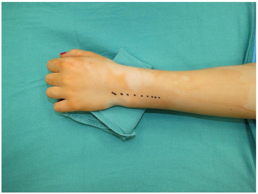 Image illustrating the surgical exposure of the Extensor Carpi Ulnaris (ECU) tendon during subsheath reconstruction
4.3. How is the Extensor Retinaculum Flap Created?
- Flap Planning: Plan a 3-cm wide flap of the extensor retinaculum.
- Flap Base: Base the flap at the ulnar border of the fifth extensor compartment.
- Radial Limit: Extend the radial limit to the radial aspect of the third compartment.
- Elevation: Carefully elevate the flap from radial to ulnar, ensuring it remains intact.
4.4. How is the Subsheath Reconstructed?
- Tendon Mobilization: Mobilize the ECU tendon from the damaged subsheath.
- Flap Passage: Pass the flap of extensor retinaculum under the ECU tendon.
- Flap Wrapping: Wrap the flap around the tendon to create a sling.
- Suturing: Suture the flap to itself using 2-0 Ethibond sutures to control the size of the new ECU tendon sheath.
- Tension Assessment: Assess the tension of the new subsheath to avoid postoperative tendonitis (if too tight) or continued instability (if too lax).
- Anchoring Sutures: Place additional sutures to anchor the flap to the adjacent extensor retinaculum for added stability.
4.5. What are Additional Considerations?
- Wrist Arthroscopy: Consider performing wrist arthroscopy during the same procedure to address other potential wrist pathologies.
- Tenosynovectomy: If tenosynovectomy is needed, it can be performed concurrently.
- TFCC Repair: Address any triangular fibrocartilage complex (TFCC) tears if present.
4.6. What Resources Does CAR-DIAGNOSTIC-TOOL.EDU.VN Provide for Surgical Guidance?
| Resource | Description | Benefits |
|---|---|---|
| Surgical Videos | Step-by-step video demonstrations of the subsheath reconstruction technique. | Provides visual guidance, improving understanding and execution of the surgical steps. |
| Anatomical Models | 3D models of the wrist anatomy highlighting the ECU tendon and subsheath. | Enhances anatomical understanding and aids in surgical planning. |
| Surgical Checklists | Comprehensive checklists to ensure all steps of the procedure are followed accurately. | Reduces the risk of errors and ensures consistency in the surgical technique. |
| Expert Consultation | Real-time consultation with experienced hand surgeons during the procedure. | Provides immediate support and helps in making critical decisions. |
| Post-operative Protocols | Detailed protocols for post-operative care and rehabilitation. | Ensures optimal recovery and reduces the risk of complications. |
By following these detailed steps and utilizing the resources provided by CAR-DIAGNOSTIC-TOOL.EDU.VN, surgeons can perform ECU subsheath reconstruction effectively, leading to improved patient outcomes and wrist stability.
5. Post-operative Rehabilitation: Regaining Wrist Function
What are the essential steps in post-operative rehabilitation for subluxing ECU tendon? Effective post-operative rehabilitation is crucial for regaining wrist function and ensuring long-term stability after ECU subsheath reconstruction. CAR-DIAGNOSTIC-TOOL.EDU.VN offers a comprehensive rehabilitation program to guide patients through the recovery process.
5.1. What is the Initial Post-operative Phase?
The initial phase focuses on protecting the surgical repair and managing pain and swelling:
- Immobilization: Place the patient in a long-arm splint with the wrist in a neutral position for 4-6 weeks.
- Elevation: Keep the hand elevated to minimize swelling.
- Pain Management: Prescribe pain medication as needed.
- Wound Care: Follow standard wound care protocols to prevent infection.
5.2. What Happens During the Intermediate Rehabilitation Phase?
This phase focuses on gradually restoring range of motion and preventing stiffness:
- Splint Removal: After 4-6 weeks, the splint is removed, and a removable wrist brace may be used.
- Range of Motion Exercises: Begin gentle range of motion exercises, including wrist flexion, extension, ulnar deviation, and radial deviation. These exercises should be performed within a pain-free range.
- Edema Control: Continue elevation and use compression bandages as needed to control edema.
- Scar Management: Start scar massage to prevent scar adhesions and improve tissue mobility.
5.3. What Exercises are Recommended During the Advanced Strengthening Phase?
This phase focuses on building strength and endurance in the wrist and forearm:
- Strengthening Exercises: Introduce progressive strengthening exercises using light weights or resistance bands. Focus on strengthening the ECU, flexor carpi ulnaris, and other wrist stabilizers.
- Proprioceptive Exercises: Incorporate proprioceptive exercises to improve wrist stability and coordination. Examples include wobble board exercises and ball toss drills.
- Functional Activities: Gradually reintroduce functional activities, such as gripping, lifting, and twisting, as tolerated.
- Sport-Specific Training: For athletes, begin sport-specific training exercises under the guidance of a physical therapist or athletic trainer.
5.4. What are the Guidelines for Returning to Activity?
Return to activity should be gradual and guided by the patient’s progress and symptoms:
- Gradual Progression: Increase activity levels gradually, avoiding sudden increases in intensity or duration.
- Pain Monitoring: Monitor for pain or swelling during and after activities. If symptoms occur, reduce the activity level and consult with a healthcare provider.
- Activity Modification: Modify activities as needed to reduce stress on the wrist.
- Protective Bracing: Consider using a wrist brace during activities to provide additional support and stability.
5.5. How Does CAR-DIAGNOSTIC-TOOL.EDU.VN Support Rehabilitation?
| Resource | Description | Benefits |
|---|---|---|
| Rehabilitation Protocols | Detailed, step-by-step rehabilitation protocols for each phase of recovery. | Ensures a structured and progressive approach to rehabilitation, maximizing functional outcomes. |
| Exercise Videos | Instructional videos demonstrating proper technique for range of motion, strengthening, and proprioceptive exercises. | Provides visual guidance, improving patient compliance and exercise effectiveness. |
| Telehealth Consultations | Remote consultations with physical therapists to monitor progress and adjust the rehabilitation program as needed. | Offers convenient access to expert guidance and support, improving patient engagement and outcomes. |
| Home Exercise Equipment Packages | Packages including resistance bands, hand grips, and other equipment needed for home exercises. | Facilitates adherence to the rehabilitation program and allows patients to continue their recovery independently. |
| Progress Tracking Tools | Tools for tracking progress, monitoring symptoms, and communicating with the healthcare team. | Enhances patient engagement and provides valuable data for optimizing the rehabilitation program. |
By following a structured rehabilitation program and utilizing the resources provided by CAR-DIAGNOSTIC-TOOL.EDU.VN, patients can achieve optimal recovery and return to their desired activities with confidence.
6. Addressing Post-Surgical Complications: Prevention and Management
What are the potential post-surgical complications and how can they be managed effectively? While ECU subsheath reconstruction is generally successful, potential complications can arise. CAR-DIAGNOSTIC-TOOL.EDU.VN provides guidance on preventing and managing these complications to ensure optimal patient outcomes.
6.1. What are Common Post-Surgical Complications?
- Infection: Surgical site infections can occur despite sterile techniques.
- Nerve Injury: The dorsal sensory branch of the ulnar nerve is at risk during surgery.
- Tendonitis: Post-operative ECU tendonitis can result from a too-tight reconstruction.
- Stiffness: Wrist stiffness can occur due to prolonged immobilization.
- Recurrent Subluxation: Although rare, the ECU tendon can subluxate again.
6.2. How Can Infections be Prevented and Managed?
- Prevention: Strict adherence to sterile surgical techniques, prophylactic antibiotics.
- Management: Antibiotics, wound care, and potential surgical debridement.
6.3. How Can Nerve Injuries be Prevented and Managed?
- Prevention: Careful surgical technique to avoid nerve damage.
- Management: Nerve repair, splinting, and physical therapy.
6.4. How Can Tendonitis be Prevented and Managed?
- Prevention: Precise sizing of the reconstructed ECU sheath.
- Management: Rest, ice, anti-inflammatory medications, and physical therapy.
6.5. How Can Stiffness be Prevented and Managed?
- Prevention: Early range of motion exercises.
- Management: Physical therapy, splinting, and, in severe cases, manipulation under anesthesia.
6.6. How Can Recurrent Subluxation be Prevented and Managed?
- Prevention: Secure and properly tensioned subsheath reconstruction.
- Management: Revision surgery.
6.7. What Support Does CAR-DIAGNOSTIC-TOOL.EDU.VN Offer for Complication Management?
| Resource | Description | Benefits |
|---|---|---|
| Complication Management Protocols | Detailed protocols for managing various post-surgical complications. | Provides a structured approach to addressing complications, improving patient outcomes. |
| Surgical Technique Videos | Videos demonstrating techniques to minimize the risk of nerve injury and ensure proper subsheath reconstruction. | Enhances surgical precision and reduces the likelihood of complications. |
| Expert Consultation | Real-time consultation with experienced surgeons for guidance on managing complex cases. | Offers immediate support and helps in making informed decisions. |
| Rehabilitation Guidelines | Specific rehabilitation guidelines for patients with complications, such as stiffness or tendonitis. | Optimizes recovery and improves functional outcomes for patients with complications. |
By understanding potential complications and utilizing the resources provided by CAR-DIAGNOSTIC-TOOL.EDU.VN, healthcare providers can effectively prevent and manage these issues, ensuring the best possible outcomes for patients undergoing ECU subsheath reconstruction.
7. Emerging Research and Techniques in ECU Tendon Management
What are the latest advancements in research and techniques for managing ECU tendon injuries? The field of ECU tendon management is continually evolving with ongoing research and the development of new techniques. CAR-DIAGNOSTIC-TOOL.EDU.VN stays at the forefront of these advancements to provide the most up-to-date information.
7.1. What are the Current Research Trends?
- Biomechanical Studies: Research focusing on the biomechanics of the ECU tendon and subsheath complex.
- Imaging Advances: Improved MRI and ultrasound techniques for better diagnosis.
- Surgical Innovations: Development of minimally invasive surgical approaches.
7.2. What Innovations are There in Imaging Techniques?
- High-Resolution Ultrasound: Provides detailed images of the ECU tendon and subsheath, allowing for dynamic assessment of tendon stability.
- 3T MRI: Offers higher resolution images compared to traditional MRI, improving the detection of subtle tendon and ligament injuries.
7.3. What are the Surgical Innovations Being Researched?
- Arthroscopic-Assisted Repair: Minimally invasive techniques for subsheath repair and reconstruction.
- Bio-Enhanced Repair: The use of biological materials to enhance tendon healing.
7.4. What are the Findings of Biomechanical Studies?
- Studies are exploring the impact of ulnar groove depth on ECU tendon stability.
- Research is investigating the effectiveness of different subsheath reconstruction techniques.
According to a cadaveric study, routine deepening of the ulnar groove did not increase the stability of the repair, suggesting that this practice may not be necessary in all cases.
7.5. How Does CAR-DIAGNOSTIC-TOOL.EDU.VN Incorporate Emerging Techniques?
| Area of Advancement | CAR-DIAGNOSTIC-TOOL.EDU.VN’s Approach | Benefits |
|---|---|---|
| Advanced Imaging | Providing access to high-resolution ultrasound and 3T MRI protocols. | Improves diagnostic accuracy and facilitates more targeted treatment planning. |
| Minimally Invasive Surgery | Offering training modules on arthroscopic-assisted repair techniques. | Reduces surgical trauma, promotes faster recovery, and improves patient outcomes. |
| Bio-Enhanced Repair | Providing information on the latest research and techniques in bio-enhanced repair. | Enhances tendon healing, reduces the risk of re-rupture, and improves long-term stability. |
| Biomechanical Insights | Incorporating biomechanical findings into surgical planning and rehabilitation protocols. | Optimizes surgical techniques and rehabilitation strategies, leading to improved functional outcomes. |
By staying informed about the latest research and incorporating emerging techniques, CAR-DIAGNOSTIC-TOOL.EDU.VN ensures that healthcare providers have access to the best available tools and knowledge for managing ECU tendon injuries.
8. Long-Term Outcomes and Patient Satisfaction: What to Expect
What are the expected long-term outcomes and patient satisfaction rates following ECU tendon surgery? Understanding the long-term outcomes and patient satisfaction rates is essential for setting realistic expectations and ensuring optimal patient care. CAR-DIAGNOSTIC-TOOL.EDU.VN provides insights into what patients can expect following ECU tendon surgery.
8.1. What are the Long-Term Results of Subsheath Reconstruction?
- Stability: Subsheath reconstruction with the extensor retinaculum flap has shown high rates of tendon stability.
- Function: Most patients regain good wrist function and can return to their previous activities.
- Pain Relief: Significant reduction in ulnar-sided wrist pain.
8.2. What do Studies Say About Patient Satisfaction?
- A single-center series published in 2020 found no recurrent subluxation after subsheath reconstruction.
- Multiple studies report high patient satisfaction rates following ECU subsheath reconstruction.
A study from Austria reported excellent satisfaction with the surgery, even in patients with persistent ECU dislocation on repeat MRI, suggesting that pain resolution may not always correlate with anatomical correction.
8.3. What Factors Influence Long-Term Outcomes?
- Surgical Technique: Precise subsheath reconstruction is essential.
- Rehabilitation: Adherence to a structured rehabilitation program.
- Patient Compliance: Following post-operative instructions and activity modifications.
- Pre-existing Conditions: Addressing any underlying wrist pathologies.
8.4. What Considerations Are Important for Patient Expectations?
- Realistic Goals: Setting realistic expectations regarding pain relief and functional recovery.
- Potential Complications: Discussing potential complications and their management.
- Rehabilitation Commitment: Emphasizing the importance of adherence to the rehabilitation program.
8.5. How Does CAR-DIAGNOSTIC-TOOL.EDU.VN Enhance Long-Term Outcomes?
| Aspect of Care | CAR-DIAGNOSTIC-TOOL.EDU.VN’s Support | Benefits |
|---|---|---|
| Surgical Excellence | Providing detailed surgical guidance and training resources. | Ensures precise subsheath reconstruction, maximizing long-term stability. |
| Rehabilitation Adherence | Offering comprehensive rehabilitation protocols, exercise videos, and telehealth consultations. | Promotes patient engagement and adherence to the rehabilitation program, improving functional outcomes. |
| Complication Management | Providing detailed protocols for preventing and managing potential complications. | Minimizes the impact of complications and optimizes patient outcomes. |
| Patient Education | Offering educational resources and support to help patients understand their condition and manage their recovery. | Empowers patients to actively participate in their care, leading to improved outcomes and satisfaction. |
By focusing on surgical excellence, rehabilitation adherence, complication management, and patient education, CAR-DIAGNOSTIC-TOOL.EDU.VN helps ensure the best possible long-term outcomes and patient satisfaction following ECU tendon surgery.
9. Optimizing ECU Tendon Healing: Nutrition and Lifestyle Factors
How can nutrition and lifestyle factors optimize ECU tendon healing post-surgery? Optimizing ECU tendon healing after surgery involves not only following medical advice and rehabilitation protocols but also paying attention to nutrition and lifestyle factors. CAR-DIAGNOSTIC-TOOL.EDU.VN emphasizes the importance of these holistic approaches for enhancing recovery.
9.1. What Role Does Nutrition Play in Tendon Healing?
- Protein: Essential for tissue repair and collagen synthesis.
- Vitamin C: Supports collagen production and acts as an antioxidant.
- Vitamin D: Important for bone health and may play a role in tendon healing.
- Omega-3 Fatty Acids: Reduce inflammation and promote tissue repair.
- Zinc: Supports wound healing and collagen synthesis.
9.2. What Foods Support Tendon Healing?
- Protein-rich foods: Lean meats, poultry, fish, eggs, dairy products, legumes, and nuts.
- Vitamin C-rich foods: Citrus fruits, berries, kiwi, bell peppers, and leafy green vegetables.
- Vitamin D-rich foods: Fatty fish, fortified dairy products, and egg yolks.
- Omega-3 fatty acid-rich foods: Fatty fish (salmon, mackerel, tuna), flaxseeds, chia seeds, and walnuts.
- Zinc-rich foods: Oysters, beef, pumpkin seeds, lentils, and whole grains.
9.3. What Lifestyle Factors Impact Healing?
- Smoking: Impairs blood flow and delays healing.
- Alcohol Consumption: Can interfere with tissue repair.
- Hydration: Essential for tissue health and nutrient transport.
- Rest and Sleep: Allow the body to repair and regenerate tissues.
9.4. What are the Recommendations for Smoking and Alcohol Consumption?
- Smoking: Quit smoking to improve blood flow and enhance healing.
- Alcohol Consumption: Limit or avoid alcohol consumption during the healing process.
9.5. What are the Guidelines for Hydration and Rest?
- Hydration: Drink plenty of water throughout the day to stay hydrated.
- Rest and Sleep: Get adequate rest and sleep to support tissue repair and regeneration.
9.6. How Does CAR-DIAGNOSTIC-TOOL.EDU.VN Support Optimized Healing?
| Aspect of Healing | CAR-DIAGNOSTIC-TOOL.EDU.VN’s Support | Benefits |
|---|---|---|
| Nutritional Guidance | Providing detailed nutritional guidelines for tendon healing. | Ensures patients consume the nutrients needed to support tissue repair and collagen synthesis. |
| Lifestyle Recommendations | Offering recommendations for lifestyle modifications to enhance healing. | Helps patients adopt habits that promote healing and avoid those that impair it. |
| Hydration Tips | Providing tips for staying adequately hydrated. | Supports tissue health and nutrient transport. |
| Sleep Hygiene Tips | Offering tips for improving sleep quality and duration. | Allows the body to repair and regenerate tissues effectively. |
By emphasizing the importance of nutrition and lifestyle factors, CAR-DIAGNOSTIC-TOOL.EDU.VN helps patients optimize ECU tendon healing and achieve the best possible outcomes.
10. FAQ: Addressing Common Questions About Subluxing ECU Tendon
Have questions about subluxing ECU tendon? Here are answers to some frequently asked questions. CAR-DIAGNOSTIC-TOOL.EDU.VN aims to provide clear and reliable information to address your concerns.
10.1. What is the Main Cause of ECU Tendon Subluxation?
The main cause is usually a tear or attenuation of the ECU subsheath, which can result from acute injuries or repetitive stress.
10.2. What are the Symptoms of Subluxing ECU Tendon?
Symptoms include ulnar-sided wrist pain, clicking or snapping sensations, and wrist instability.
10.3. How is ECU Tendon Subluxation Diagnosed?
Diagnosis involves physical examination techniques, such as the ECU synergy and subluxation tests, as well as imaging studies like ultrasound and MRI.
10.4. Can ECU Tendon Subluxation Heal on Its Own?
Minor subsheath tears may heal with conservative management, but significant tears often require surgical intervention.
10.5. What is the Conservative Treatment for ECU Tendon Subluxation?
Conservative treatment includes immobilization, physical therapy, and activity modification.
10.6. When is Surgery Necessary for ECU Tendon Subluxation?
Surgery is considered when conservative measures fail to provide relief or in cases of chronic instability.
10.7. What are the Surgical Options for ECU Tendon Subluxation?
Surgical options include primary subsheath repair and subsheath reconstruction.
10.8. What is the Recovery Time After ECU Tendon Surgery?
Recovery typically involves 4-6 weeks of immobilization followed by a structured rehabilitation program.
10.9. What Activities Should be Avoided After ECU Tendon Surgery?
Avoid strenuous physical activities for three months after surgery.
10.10. What Resources Does CAR-DIAGNOSTIC-TOOL.EDU.VN Offer for ECU Tendon Management?
CAR-DIAGNOSTIC-TOOL.EDU.VN offers diagnostic tools, surgical guidance, rehabilitation protocols, and expert consultation to support ECU tendon management.
With these answers to frequently asked questions, CAR-DIAGNOSTIC-TOOL.EDU.VN aims to provide valuable information and support for individuals seeking to understand and manage subluxing ECU tendon.
Are you ready to take the next step in diagnosing and treating subluxing ECU tendon effectively? Contact CAR-DI